(一)目前的多重检测技术主要是检测显色染料、金属同位素、荧光基团等发出的信号来达到检测标志物的目的。各个mIHC/IF平台的工作原理图(Diagram showing mechanism of each of the mIHC/IF platform)如图1所示:
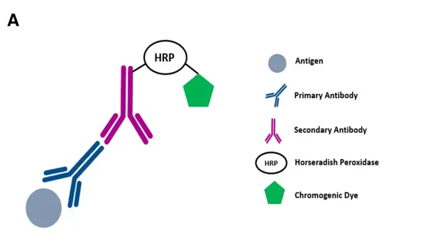
图1A.DISCOVERY ULTRA system: after primary antibody incubation, a secondary antibody labelled with HRP is introduced. The HRP is reacted with an appropriate substrate bound to a chromogenic dye, leading to the precipitation of insoluble, coloured precipitates at the site where the antigens are found.
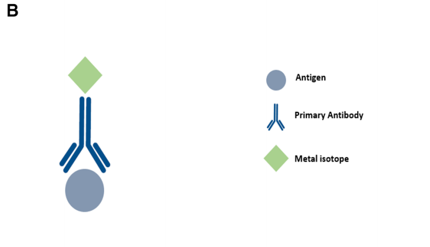
图1B.Metal-based IHC techniques such as IMC and MIBI:a primary antibody bound to the target antigen is tagged with a metal isotope of known molecular mass. Analysis is carried out using mass spectrometry in MIBI and laser ablation coupled to mass cytometry in IMC.
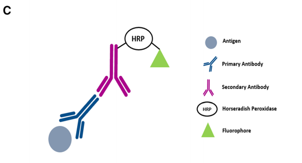
图1C.Vectra: after primary antibody incubation, a secondary antibody labelled with HRP is introduced. A fluorophore-conjugated tyramide molecule serves as the substrate for HRP, resulting in an antigen-associated fluorescence signal.
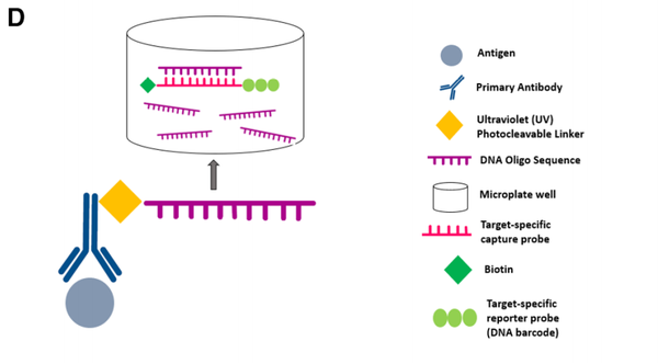
图1D.Nanostring’s DSP: the target antigen will bind the primary antibody which is coupled to a photo cleavable oligonucleotide tag. UV light is used to cleave the oligonucleotide tags and is collected using a micro capillary tube and stored in a microplate well. The oligonucleotide tags will bind to the reporter probe via the target-specific capture probe. Reporter probes are imaged and counted by the nCounter analysis system.
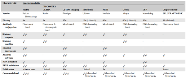
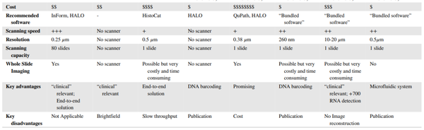
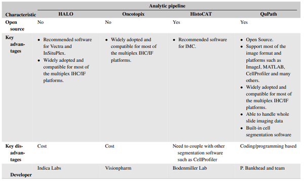
(二)现有的多重检测技术可同时检测5-40+标志物,不同成像平台的比较(Overview and comparison of the different imaging modalities)如表1所示:
(三)Vectra, Chipcytometry, or DISCOVERY ULTRA平台mIHC/IF图像示例(Representative mIHC/IF images captured through the Vectra, Chipcytometry, or DISCOVERY ULTRA imaging system):
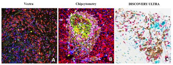
图2. Representative mIHC/IF images captured through the Vectra, Chipcytometry, or DISCOVERY ULTRA imaging system.
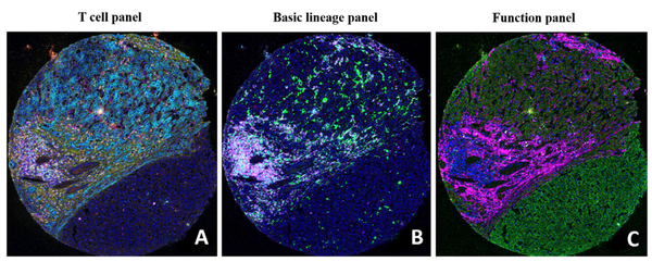
(六)Ultivue’s InSituPlex可使用普通的荧光显微镜成像,不需要配置昂贵的显微镜,因此可在大部分实验室开展检测。图4为人体组织样本Ultivue’s InSituPlex图像示例(Representative Ultivue’s InSituPlex images of human tissue samples labelled with CD8 (green), CD68 (yellow), PD-L1 (red) and CK/Sox10 (cyan)):
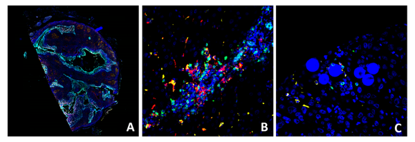
图4. Representative Ultivue’s InSituPlex images of human tissue samples labelled with CD8 (green), CD68 (yellow), PD-L1 (red) and CK/Sox10(cyan). Whole slide imaging of tonsil section (A), high magnification view ofHCC (B), and radioembolization-treated HCC (C, Y-90 visible as microspheres).
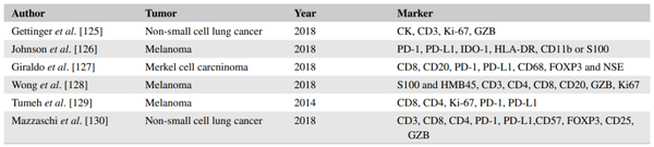
(七)目前已研究过多种mIHC/IF Panel,通过同时分析多个标志物来预测患者接受PD-1/PD-L1抑制剂治疗的应答反应。Meta分析中使用mIHC/IF技术的文献清单(List of papers using mIHC/IF in the meta-analysis)如下:
杰论

综上所述,mIHC/IF在肿瘤免疫治疗领域具有广阔的应用前景。与传统的IHC只能检测一个标志物不同,mIHC/IF能够在单个组织切片中检测多个标志物,同时提供有关细胞组成和空间排列的全面信息,使我们能够更深入地了解癌症的发病机制和对免疫治疗的反应。同时mIHC/IF技术处理的组织样本可以长期保存,供进一步研究使用。但是,这类技术的检测成本及实用性方面,仍然是一个值得关注的问题。
迈杰转化医学作为国内精准诊断整体解决方案的领导者,致力于解决精准医疗药物研发及患者用药痛点,围绕生物标志物研究、伴随诊断开发,建立了完善的核酸组学、蛋白组学、细胞组学技术平台。我们拥有国内领先的IHC检测平台,配备有Leica Bond Max、Ventana BenchMark、Dako Autostainer Link48三大进口自动化平台,以及用于mIHC检测的Leica Bond RX、PerkinElmerVectra3 System平台,在mIHC检测方法学开发及验证方面积累了丰富的经验,如三标四色的Panel(CD4, CD8, PD-L1 ),及五标六色的Panel(CD4, CD8, CD163, PD-L1, Pan-Keratin)等,如有mIHC检测方法开发及服务需求,请联系迈杰转化医学商务部(邮箱:MARKETING@MEDxTMC.com)。
文献链接
Overview of multiplex immunohistochemistry/immunofluorescence techniques in the era of cancer immunotherapy - Tan - 2020 - Cancer Communications - Wiley Online Library(https://onlinelibrary.wiley.com/doi/10.1002/cac2.12023
▎迈杰转化医学市场部团队编辑

 关注微信公众号

 English
有限公司")